
Timisoara_Med 2022, 2022(2), 1; doi:10.35995/tmj20220201
Article
One-Year Follow-up Study on Minimally Invasive Treatment of Cariogenic or Non-cariogenic White Spot Lesions
1
Discipline of Preventive, Community Dentistry and Oral Health, Faculty of Dental Medicine, “Victor Babeș” University of Medicine and Pharmacy, 2 Eftimie Murgu Sq., 300041 Timișoara, Romania; nituraluca30@gmail.com (R.L.N.); pascaioanagiorgiana@gmail.com (I.G.P.); damian.lia-raluca@umft.ro (L.R.D.); dumitrescu.ramona@umft.ro (R.C.D.); roancea@umft.ro (R.O.)
2
Department of Oral Health and Community Dentistry, Faculty of Dental Medicine, “Carol Davila” University of Medicine and Pharmacy, 17-23 Calea Plevnei Street, 010221 Bucharest, Romania
*
Correspondence: ruxandra.sfeatcu@gmail.com
How to cite: Nițu, R.L.; Sfeatcu, R.; Pasca, I.G.; Damian, L.R.; Dumitrescu, R.C.; Oancea, R. One-Year Follow-up Study on Minimally Invasive Treatment of Cariogenic or Non-cariogenic White Spot Lesions. Timisoara Med. 2022, 2022 (2), 1; doi:10.35995/tmj20220201.
Received: 11 April 2022 / Accepted: 3 June 2022 / Published: 30 July 2022
Abstract
:(1) Background: The current practice in dental medicine aims to perform non-invasive treatments, which preserve as many hard tissues as possible. This trend is based on the final appearance, which should be as aesthetic and as close to natural teeth as possible. The use of Icon DMG can be considered revolutionary in the treatment of white spot lesions (WSLs) caused by various factors. (2) Methods: A diagnosis was established after clinical inspection, transillumination, or based on the patient’s medical history. Icon infiltration was followed according to the producer’s indications. Chromatic stability was assessed by two examinators with Vitapan 3D Master, and gum health improvement was assessed using the gingival index (GI). (3) Results: Ten dental units were observed for one year and showed chromatic stability. Gum health was improved. No side effects occurred. (4) Conclusions: An easy, non-invasive technique and painless treatment are the desired results from the use and implementation of resin in current practice. Following the infiltration protocol offers the desired aesthetic results.
Keywords:
white spot lesions (WSLs); resin infiltration; chromatic stability; gum healthIntroduction
White spot lesions (WSLs) are the clinical manifestation of an initial carious lesion, and they become visible due to the different refractive indexes of healthy and demineralized enamel. They are characterized by an opaque white area surrounded by healthy enamel. These are the first signs of teeth demineralization, and they occur on the smooth surfaces of the teeth. The enamel loses its translucency, which is directly connected to the degree of mineralization. Initially, pores are created between the enamel prisms, and as they develop, a porous structure is established [1]. For the proper clinical detection of WSLs, dental surfaces should be clean and plaque-free. Drying the surfaces of teeth has two functions: the removal of saliva—which can camouflage a lesion—and the dehydration of a white spot lesion. If the lesion is visible on damp surfaces, it indicates a demineralization of more than half the thickness of the enamel, even reaching the outer third of dentin [2]. WSLs can also occur as a result of various anomalies in the structure of the enamel. Structural abnormalities result from the disruption of the process of histodifferentiation, apposition and/or mineralization. Enamel defects are due to hypoplasia or hypocalcification, which may be generically inherited (imperfect amelogenesis) or may be caused by the environment (local or systemic causes) [3]. For example, one isolated WSL on the smooth surface of a tooth may be caused by a traumatic episode experienced by the temporary tooth, while many WSLs—white and/or brown—are caused by dental fluorosis; one WSL in the cervical third surrounded by brown margins is a sign of postorthodontic WSL [4].
The concept of resin infiltration is to infuse inside the pores of the enamel through capillarity in order to seal pathways to prevent further acid penetration, arresting the evolution of dental caries. This technique aims to create a diffusion barrier inside the lesion, not just on its surface [5].
Resin infiltration has made it possible to stop the evolution of dental caries and mask various structural defects, perceived as inaesthetic by the patient. This product fits perfectly into the concept of minimally invasive dentistry, reconciling even the most anxious patients by having a number of advantages, including arresting the carious lesion, being a rapid treatment following the concept of single-visit dentistry, preserving dental tissues, and being a painless treatment with outstanding aesthetic results.
This study aims to monitor the evolution and chromatic stability of infiltrated WSLs on smooth surfaces and the improvement of gingival health at one, six, nine, and twelve months after the icon infiltration procedure.
Material and Methods
This study began in November 2019, after the consent of the Ethics Commission of the Faculty of Dentistry Timisoara, Romania, was obtained, and was conducted up until May 2021. Each subject voluntarily confirmed their participation in this study and signed an informed consent form.
Initially, a group of 15 patients were registered, but only seven met the inclusion criteria (18–25 years old; WSL on smooth tooth surfaces). There was a total of 21 teeth that underwent the infiltration protocol. Of the 7 eligible subjects, 3 (2 males, 1 female) came to regular check-ups at periods of 1, 6, 9 and 12 months, adding up to 11 teeth that were followed up; the diagnosis was established after patients’ in-depth medical histories were obtained, where it was found out that one patient took fluoride supplements daily until the age of 8 years. The other one underwent orthodontic treatment for 2 years; she presented herself in the office with a WSL at 3 months after the treatment ended. The exclusion criteria were dental units presenting cavities and/or previous fillings, temporary teeth, underage patients, and smokers.
Icon comes in two distinct forms—for smooth or proximal surfaces, wherefrom we chose the Vestibular version. A thorough cleaning of the tooth surface is mandatory by applying 15% Hydrochloric acid (Icon Etch) for 2 min, which needs to be reactivated with an applicator every 1 min. We used 15% HCl as it proved to be superior than the 37% Orthophosphoric acid in removing the mineralized area and produces a penetration of 58 µm, while the latter—only 25 µm [5].
The ethanol wet-bonding technique is designed to dry the lesion out by applying 99% ethanol (Icon Dry) for 30 s, followed by drying the surface. Ethanol facilitates the penetration of hydrophobic monomers into the demineralized lesion. This way, the water in the demineralized collagen matrix is replaced with ethanol of increasing concentration, allowing the latter to enter the collagen matrix without causing the further contraction of the interfibrillar space [6,7].
Icon Infiltrant, composed of triethylene glycol dimethacrylate (TEGDMA), was applied to the surface of the lesion and requires 3 min of action. The excess was removed and then light-cured. In order to compensate for polymerization shrinkage, this step was repeated once more by applying the low-viscosity resin for another minute. In the end, the infiltrated surfaces were light-cured and finished [8,9].
Subsequently, periodic check-ups were performed at 1, 6, 9, and 12 months. Both at the beginning of and during the periodic controls, the chromatic evolution of the lesions was registered with Vitapan 3D Master. Teeth color was assessed by two independent examinators.
Regarding periodontal examination, the parameter measured for assessing gingival inflammation was the initial and final gingival index (GI). GI was assessed at 4 sites (mesial, buccal, distal, and lingual/palatal) of each tooth that presented WSLs in the cervical third of the buccal surface: a score of 0—normal; a score of 1—mild gingival inflammation without bleeding on probing; a score of 2—moderate inflammation with bleeding on probing; a score of 3—severe inflammation with a tendency to spontaneously bleed) [10].
Results
The 11 infiltrated dental units were followed for one year. Even though the initial result—right after the treatment—was partially successful, one month later, the appearance improved because the teeth were given enough time to rehydrate, their color stabilized, and the gingival margin healed.
Possible chemical lesions of the gingival margin were followed (Table 1), as was the chromatic stability of the resin. It was found that, in addition to the persistent stability, there was also a chameleon effect over time (Figure 1). One patient showed mild sensibility for three days after the procedure.
The color of the teeth was determined during the first session, after one month and after one year with Vitapan 3D Master. There was no difference regarding the shade of the infiltrated dental units (Table 1).
Regarding GI, there was an improvement in the score after infiltration near the gingival margins. On clinical examination, the initially inflamed gingival margin and pinkish-red color healed after one month (Table 2).
Discussion
Recently, there has been a dramatic change in the management of dental caries—from the traditional restorative treatment to a preventive one, called noninvasive or minimally invasive [11]. Caries are initially characterized by a loss of minerals, which results from the opacification of the enamel. This phenomenon is visible due to the alteration of the refractive index of that area [12].
Particular attention was paid to the non-invasive treatment of incipient enamel lesions, treating them by remineralization with casein phopshopeptide-amporphus calcium phosphate (CPP-ACP). However, it has been shown that remineralization with toothpaste containing CPP-ACP is not always successful, as it requires patient cooperation and giving up bad cariogenic habits, and they often abandon treatment, compared with the result we obtained with resin infiltration. It has been found that the porous structure of enamel is beneficial for the diffusion of dissolved acids and minerals [13]. This concept has been modified and marketed in Germany for the management of initial lesions on the buccal/lingual/proximal surfaces by infiltrating them with a low-density resin.
There are two aspects to consider when it comes to treatment for a patient suffering from WSLs. One is preventing caries from becoming cavitary when the remineralizing power of saliva is failing, and another one is masking the aesthetic damage caused by WSLs in the front area. WSLs on front teeth should be treated for two reasons: to improve the appearance of a patient’s smile and to prevent cavities.
Munoz MA et al. noted that fluorotic and hypomineralized enamels have significantly reduced mineral content, similar to an incipient carious lesion. Therefore, the infiltration technique is recommended for masking the unpleasant white spots, as shown in this study [14].
Another important aspect is the protection of gingival margins. Like CRG Torres and AB Borges, we believed that the soft tissue should not come in contact with HCl because it has an aggressive behavior on organic tissues. Especially in cases of fluorosis, as seen in this study, the lesions can be spread on the entire buccal surface, including the cervical third [15]. This is why it was decided to isolate them with rubberdam, floss ligatures, and liquid dam. This type of isolation has the role of retracting the gingival tissue, revealing the entire cervical area, which can be infiltrated the right way. Otherwise, WSLs could persist in this critical area and will compromise the final result. On the other hand, infiltrating the WSLs improved gum health and the gingival margins were not affected.
The topic of postorthodontic WSLs has been approached by several researchers regarding the ability of the product to mask any lesions, concluding that they can be treated using the same protocol—with the same results. A six-month randomized study by Knosel et al. verified the chromatic stability of the infiltrant [16]. As shown in this study, they observed a perfect integration of the color and brightness of the infiltrated WSLs compared to the healthy peripheral enamel [16]. Fixed orthodontic appliances facilitate the accumulation of dental plaque and can lead to WSLs [17]. Many studies concluded that WSLs occur in the first few weeks of fixed orthodontics therapy [18,19], reaching a prevalence of up to 40% during the first half of the year [19].
One study conducted by Choi EM, Park BY, and Noh HJ presents a systematic review to assess the clinical impact of sending periodic notifications to the patients undergoing orthodontic treatment for improving dental hygiene [20]. Nine out of eleven studies registered improved oral hygiene by obtaining decreased scores of the oral hygiene indices, periodontal indices, and lowering the appearances of WSLs [21,22,23,24,25,26,27,28,29].
Conclusions
In this study, we assessed the accuracy of Icon DMG, the implications of resin infiltration for gum health, and its chromatic stability over time by targeting WSLs of different etiologies. Following this study, it can be concluded that the infiltration technique is easy to carry out, its efficiency is increased for various etiologies, the working time is significantly shortened, the resin is dimensionally and chromatically stable and gingival health improves. This procedure is painless and the postprocedural sensitivity is temporary, while the results are particularly aesthetic.
Author Contributions
Conceptualization, R.L.N., R.S. and R.O.; Methodology, R.L.N., R.S. and R.O.; Validation, I.G.P., R.L.D. and R.C.D.; Formal Analysis, R.L.N., R.S. and R.O.; Investigation, R.L.N.; Resources, R.L.N.; Data Curation, I.G.P., R.L.D. and R.C.D.; Writing—Original Draft Preparation, R.L.N.; Writing—Review and Editing, R.L.N., I.G.P., R.L.D. and R.C.D. Supervision, R.S. and R.O.
Funding
This research received no external funding.
Conflicts of Interest
The authors declare no conflict of interest.
References
- Summit, J.; Robbins, J.; Schwartz, R. Fundamentals of Operative Dentistry: A Contemporary Approach, 2nd ed.; Quintessence books: Chicago, IL, USA, 2001; pp. 1–8. [Google Scholar]
- Heymann, H.; Swift, E.J.; Ritter, A. Sturdevant’s Art and Science of Operative Dentistry, 6th ed.; Elsevier/Mosby: Chicago, IL, USA, 2013; pp. 220–225. [Google Scholar]
- Nowak, A.; McTigue, D.; Casamassimo, P.; Fields, H. Pediatric Dentistry: Infancy through Adolescence, 5th ed.; Elsevier: Amsterdam, The Netherlands, 2012; pp. 58–61. [Google Scholar]
- Weisrock, G.; Terrer, E.; Couderc, G.; Koubi, S.; Levallois, B.; Manton, D.; Tassery, H. Naturally aethetic restorations and minimally invasive dentistry. J. Min. Intervation Dent. 2011, 4, 23–30. [Google Scholar]
- Manoharan, V.; Kumar, S.A.; Arumugam, S.B.; Anand, V.; Krishnamoorthy, S.; Methippara, J.J. Is Resin Infiltration a Microinvasive Approach to White Lesions of Calcified Tooth Structures? A Systemic Review. Int. J. Clin. Pediatric Dent. 2019, 12, 53–58. [Google Scholar]
- De Barros, L.; Apolonio, F.M.; Loguercio, A.D.; De Saboia, V. Resin-Dentin Bonds of Etch-and-Rinse Adhesives to Alcohol-saturated Acid-etched Dentin. J. Adhes. Dent. 2013, 15, 333–340. [Google Scholar] [PubMed]
- Li, F.; Liu, X.-Y.; Zhang, L.; Kang, J.-J.; Chen, J.-H. Ethanol-wet Bonding Technique May Enhance the Bonding Performance of Contemporary Etch-and-Rinse Dental Adhesives. J. Adhes. Dent. 2012, 14, 113–120. [Google Scholar] [PubMed]
- Lasfargues, J.J.; Bonte, E.; Guerrieri, A.; Fezzani, L. Minimal intervention dentistry: Part 6. Caries inhibition by resin infiltration. Br. Dent. J. 2013, 214, 53–59. [Google Scholar] [CrossRef] [PubMed]
- Kim, S.; Kim, E.-Y.; Jeong, T.-S.; Kim, J.-W. The evaluation of resin infiltration for masking labial enamel white spot lesions. Int. J. Paediatr. Dent. 2011, 21, 241–248. [Google Scholar] [CrossRef] [PubMed]
- Scott, B. Schwartz JRCHF. 31—Examination, Diagnosis and Treatment Planning. In Pediatric Dentistry, 6th ed.; Arthur, J., Nowak, J., Eds.; Elsevier: Amsterdam, The Netherlands, 2019; pp. 419–454. [Google Scholar]
- Holmgren, C.; Domejean, S.; Ducamp, R.; Léger, S. Resin infiltration of non-cavited caries lesions: A systematic review. Med. Princ. Pract. 2015, 24, 216–221. [Google Scholar]
- Featherstone, J.D. The caries balance: The basis for caries managemnt by risk assessment. Oral. Health Prev. Dent. 2004, 2, 265–269. [Google Scholar]
- Torres CR, G.; Rosa PC, F.; Ferreira, N.S.; Borges, A. Effect of Caries Infiltration technique and fluoride therapy on microhardness of enamel carious lesions. Oper. Dent. 2012, 37, 363–369. [Google Scholar] [CrossRef]
- Muñoz, M.A.; Arana-Gordillo, L.A.; Gomes, G.M.; Gomes, O.M.; Bombarda, N.H.C.; Reis, A.; Loguercio, A.D. Alternative esthetic management of fluorosis and hypoplasia stains: Blending effect obtained with resin infiltration techniques. J. Esthet. Restor. Dent. 2013, 25, 32–33. [Google Scholar] [CrossRef]
- Torres CR, G.; Borges, A.B. Color masking of developmental enamel defects: A case series. Oper. Dent. 2015, 40, 25–33. [Google Scholar] [CrossRef]
- Knösel, M.; Eckstein, A.; Helms, H.J. Durability of esthetic improvement following Icon resin infiltration of multibracket-induces white sport lesions compared with no therapy over 6 months: A single-center, split-mouth, randomized clinical trial. Am. J. Orthod. Dentofac. Ortop. 2013, 144, 86–96. [Google Scholar] [CrossRef]
- Ren, Y.; Jongsma, M.A.; Mei, L.; van der Mei, H.C.; Busscher, H.J. Orthodontic treatment with fixed appliances and biofilm formation–a potential public health threat? Clin. Oral. InvestIG. 2014, 18, 1711–1718. [Google Scholar] [CrossRef]
- Tufekci, E.; Dixon, J.S.; Gunsolley, J.C.; Lindauer, S.J. Prevalence of white spot lesions during orthodontic treatment with fixed appliances. Angle Orthod. 2011, 81, 206–210. [Google Scholar] [CrossRef]
- Lucchese, A.; Gherlone, E. Prevalence of white-spot lesions before and during orthodontic treatment with fixed appliances. Eur. J. Orthod. 2013, 35, 664–668. [Google Scholar] [CrossRef]
- Choi, E.; Park, B.; Noh, H. Efficacy of mobile health care in patients undergoing fixed orthodontic treatment: A systematic review. Int. J. Dent. Hyg. 2020, 19, 29–38. [Google Scholar] [CrossRef]
- Zotti, F.; Dalessandri, D.; Salgarello, S.; Piancino, M.; Bonetti, S.; Visconti, L.; Paganelli, C. Usefulness of an app in improving oral hygiene compliance in adolescent orthodontic patients. Angle Orthod. 2016, 86, 101–107. [Google Scholar] [CrossRef]
- Alkadhi, O.H.; Zahid, M.N.; Almanea, R.S.; Althaqeb, H.K.; Alharbi, T.H.; Ajwa, N.M. The effect of using mobile applications for improving oral hygiene in patients with orthodontic fixed appliances: A randomised controlled trial. J. Orthod. 2017, 44, 157–163. [Google Scholar] [CrossRef]
- Li, X.; Xu, Z.-R.; Tang, N.; Ye, C.; Zhu, X.-L.; Zhou, T.; Zhao, Z.-H. Effect of intervention using a messaging app on compliance and duration of treatment in orthodontic patients. Clin. Oral Investig. 2015, 20, 1849–1859. [Google Scholar] [CrossRef]
- Bowen, T.B.; Rinchuse, D.J.; Zullo, T.; DeMaria, M.E. The influence of text messaging on oral hygiene effectiveness. Angle Orthod. 2014, 85, 543–548. [Google Scholar] [CrossRef]
- Cozzani, M.; Ragazzini, G.; Delucchi, A.; Mutinelli, S.; Barreca, C.; Rinchuse, D.J.; Servetto, R.; Piras, V. Oral hygiene compliance in orthodontic patients: A randomized controlled study on the effects of a post-treatment communication. Prog. Orthod. 2016, 17, 14. [Google Scholar] [CrossRef] [PubMed]
- Eppright, M.; Shroff, B.; Best, A.; Barcoma, E.; Lindauer, S.J. Influence of active reminders on oral hygiene compliance in orthodontic patients. Angle Orthod. 2014, 84, 208–213. [Google Scholar] [CrossRef] [PubMed]
- Kumar, G.S.; Kashyap, A.; Raghav, S.; Bhardwaj, R.; Singh, A.; Guram, G. Role of text message reminder on oral hygiene maintenance of orthodontic patients. J. Contemp. Dent. Pract. 2018, 19, 98–101. [Google Scholar] [PubMed]
- Al-Anezi, S.A.; Harradine, N.W.T. Quantifying plaque during orthodontic treatment. Angle Orthod. 2012, 82, 748–753. [Google Scholar] [CrossRef]
- Huang, J.; Yao, Y.; Jiang, J.; Li, C. Effects of motivational methods on oral hygiene of orthodontic patients: A systematic review and meta-analysis. Medicine 2018, 97, e13182. [Google Scholar] [CrossRef]
Figure 1.
Initial, intermediary and final aspects of the followed infiltrated teeth. (a) The initial situation; (b) right after the infiltration; (c) one year follow-up.
Figure 1.
Initial, intermediary and final aspects of the followed infiltrated teeth. (a) The initial situation; (b) right after the infiltration; (c) one year follow-up.
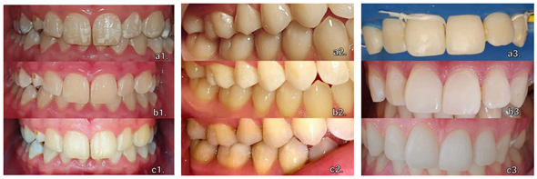

Table 1.
Initial situation and chromatic stability.
| WSL | Number of Teeth | Etching Time (min) | Tooth Color—Initial | Tooth Color—Final |
|---|---|---|---|---|
| Dental fluorosis | 8 | 6 min | 2L 1.5 | 2L 1.5 |
| WSL after orthodontic treatment | 1 | 3 min | 2R | 2R |
| Post-traumatic WSLs | 2 | 6 min | 3M 2 | 3M 2 |
Table 2.
Gingival index for assessing gingival inflammation.
| WSL | GI Initial Score | GI Final Score | |||||||||||||
|---|---|---|---|---|---|---|---|---|---|---|---|---|---|---|---|
| Dental fluorosis | Score | 2 | 1 | 1 | 1 | 2 | 1 | 0 | 1 | 0 | 1 | 0 | 1 | 0 | 0 |
| Average | 1.142 | 0.28 | |||||||||||||
© 2022, Copyright by the authors Licensed as an open access article using a CC BY 4.0 license